Location: Home >> Detail
Adv Geriatr Med Res. 2026;8(3):e260019. https://doi.org/10.20900/agmr20260019
 ,
Nina Daneshvar 1 ,
Bryan Godfrey 1 ,
Jennifer Muise 1 ,
Tess Thompson 2,3 ,
Denisé Dews 1
,
Nina Daneshvar 1 ,
Bryan Godfrey 1 ,
Jennifer Muise 1 ,
Tess Thompson 2,3 ,
Denisé Dews 1
1
2
3
*
Background: Caregivers of older adults in rural settings face distinct challenges shaped by environmental factors, including longer travel distances, limited support networks, and fewer resources. Addressing these unmet needs is critical to reducing burnout and negative health outcomes. Methods: We surveyed care managers to compare rural caregivers’ needs with urban caregivers. A convenience sample of care managers completed a survey developed by geriatrics professionals. The survey included multiple-choice and seven optional open-ended questions. Participants were recruited via email and word of mouth (March–June 2025). Inclusion criteria required completion of more than half the survey, providing a practice ZIP code, and employment in North Carolina. Quantitative data were analyzed descriptively; qualitative responses were analyzed by a hybrid deductive-inductive thematic approach. Results: The participation rate was 16% (62/387) before exclusions. The final sample included 36 participants, 89% of whom completed the entire survey. Most were aged 25–34 (38.9%), held a master’s degree (55.6%), had 1–5 years of experience (52.8%), and practiced in rural settings (50%). A total of 89 open-ended responses were analyzed; six responses were reported as “Unsure” and not included in the theme development. Two themes emerged: (1) there are fewer resources for caregivers in rural compared with urban communities, and (2) care managers in rural communities face additional barriers beyond limited resources. Conclusion: Rural caregivers, and the care managers who support them, face distinct challenges. This exploratory work informs future research to expand to other regions, standardize rural definitions in research, and develop and evaluate system-level interventions to better support both caregivers and care managers.
There are over 24 million informal or unpaid caregivers for older adults (65+ years) in the United States [1]. Rural areas have higher proportions of informal caregivers than in urban areas, with caregiving duties being filled by families and loved ones [2]. Informal caregivers face significant stressors related to caregiving requirements, which are associated with poor health, financial strain, and poor quality of life [3,4]. While informal caregiving patterns vary across the United States, rural living is associated with higher caregiver strain and worse health outcomes due to the limited resources for caregiver support [2,5–7]. Additionally, rural areas may present unique neighborhood planning challenges, such as lower walkability, that can reduce older adults’ sense of community and perceived social support, potentially placing additional strain on older adult-caregiver relationships and increasing support needs [8]. However, there is little previous research on understanding the caregiver-care recipient dyad needs in rural areas and the needs of the care managers that support them.
The clinical care manager holds an important role, providing support for informal caregivers and the older adult patients by assisting with resource acquisition, navigating the healthcare system, and coordination of care. The care manager, often a social worker or nurse by training, provides information about support services in the community to the dyad. The care manager often interfaces with the caregiver due to barriers to interactions with the older adult themselves (e.g., cognitive impairment, severe chronic illness). However, rural areas have fewer caregiver support services such as home health, meal delivery, companion care, including care coordination by a care manager [9–12]. Therefore, care managers can struggle to provide resources to address the unmet needs of informal caregivers in rural areas. Care managers have unique insight into the unmet needs of informal caregivers, often being the first line of contact for the caregiver when searching for care support. Exploring the care manager perspective is important for better understanding the gaps in the healthcare system and how to effectively address them in under-resourced areas. This exploratory study aims to describe and compare the needs of informal caregivers of older adults in rural and urban areas from the perspective of care managers working in these settings.
Participants were eligible for the study if they self-identified as a care manager, licensed social worker, and or nurse navigator working with older adult populations (65 years and older) and their caregivers. Participants were ineligible for the study if they completed less than 50% of the survey and or did not provide ZIP code of practice location. Survey participation was anonymous.
At the authors’ home academic institution, HS spoke to clinical groups via online video platform to notify them of the survey and provided survey links and a QR code during one encounter (February 2025). To minimize response bias at the home academic institution, invitations to participate were sent via listservs and surveys responses were collected anonymously. All other survey recruitment was conducted via email to national social work organizations, social work departments in hospitals and academic institutions via a central email announcement, and individual social work emails meeting criteria identifiable online. The initial intention was for this to be a nationwide study; however, due to n = 5 of the sample being outside of North Carolina, they were not included in the analysis, as n = 2 did not meet the 50% survey completion requirement. Of the total 387 email addresses contacted, 62 responded (16%), with n = 36 meeting study criteria.
This was considered an exempt study by the University of North Carolina-Chapel Hill Institutional Review Board (IRB #24-3130). At the beginning of the survey, participants were given information about the study, self-reported eligibility information, and were considered to have consented if they indicated they were willing to participate.
Survey DevelopmentSurvey questions were developed by an interdisciplinary team comprised of four social workers, one geriatrician, and one medical student. Question content was based on the team’s expert opinion about types of and intensity of support needed by older adults (65 years and older) and their caregivers in rural versus urban communities. Table S1 shows the survey items. Participants reported demographic information and years of employment.
Rural DefinitionRural versus urban areas of practice were determined using the “Am I Rural” website (https://www.ruralhealthinfo.org/am-i-rural#/), which provides various definitions of rurality based on ZIP code [13]. Based on the provided ZIP code, we considered a participant to practice in a rural area if they were eligible for the Centers for Medicare and Medicaid Services Rural Clinics Program, due to importance of clinician knowledge of this designation in terms of governmental support services. Because there is no gold standard definition of rural, we opted for a ZIP code-based definition in order to easily define rural and examine whether the participant’s perspective of their practice location (rural vs. urban) was correct. A proportion of the results are organized rural and urban practice location to see if practice location yields any observation patterns.
Statistical AnalysisUnivariate and bivariate statistical analyses were conducted to describe the sample.
The Chi-squared test was used to examine differences in categorical responses between rural and urban practitioners. For responses with expected cell counts < 5, we used Fisher’s exact test. We performed a sensitivity analysis comparing care manager quantitative responses based on self-reported rural and urban practices areas. Because themes identified from the free-text responses were consistent across rural and urban groups, a sensitivity analysis was not performed for the qualitative data.
Free-text responses were analyzed using a hybrid deductive-inductive thematic approach. Informed by the study objectives, HS identified preliminary themes prior to reviewing the seven open-ended survey questions. HS refined the codebook after the initial review to best describe the responses, subsequently consolidating overlapping themes into a preliminary coding framework. ND independently reviewed the coding framework and associated responses, and discrepancies in theme identification or interpretation were resolved through discussion until consensus was reached. Themes were then refined to ensure they accurately reflected participant responses. Consistent with Braun and Clarke’s guidance on thematic analysis, the findings are presented as a descriptive summary of recurring patterns supported by representative participant quotations [14]. Given the brief, optional nature of the survey responses, the objective was to identify common themes rather than achieve formal theoretical saturation. Therefore, the adequacy of the qualitative data was considered using Malterud et al.’s concept of information power, whereby focused study aims and a relatively specific participant population can support meaningful thematic interpretation despite a modest number of responses [15].
Stata 19 (College Station, Texas) and Dedoose 10.0.34 (Los Angeles, California) were used to organize and analyze quantitative and qualitative samples, respectively.
Descriptive statistics of the sample are displayed in Table 1. There were a total of 36 participants and 89.0% completed the entire survey and n = 4 completed 53%–70%. The majority of participants completed a master’s degree level of education and practiced in a hospital setting with 1–5 years’ experience. Many (n = 12) of the participants described their job role as “other,” with an assortment of care management/coordination, administration, and educational positions listed. Approximately 50% of participants reported practicing in a rural area, and approximately 55% of participants correctly reported their practice ZIP code as rural versus urban. Additional quantitative results (by question) are presented in Table S2. Most notably, needs were thought to be similar for rural and urban caregivers, except for financial support for caregivers--respondents believed that financial support was a greater need for caregivers in rural (94.7%) compared to urban areas (62.5%) (p = 0.03). Table S3 shows the sensitivity analysis for comparing perceptions of care managers based on self-reported geographical area of practice. Care managers from self-reported areas felt that caregivers from rural areas had more requests than urban caregivers (p = 0.005). Otherwise, similar to the initial analysis in Table S2, results were non-significant.
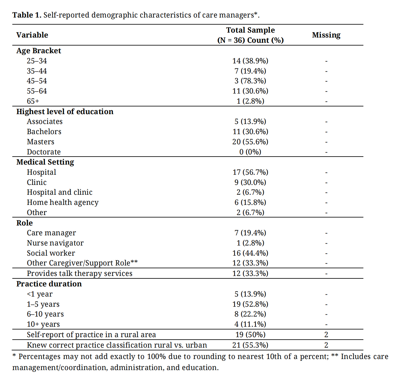 Table 1. Self-reported demographic characteristics of care managers*.
Responses to open-ended questions in Table S1 were analyzed to identify major inductive themes, with illustrative quotations integrated into the narrative and additional stand-alone quotations provided in Table 2. Free-response content substantially overlapped between urban and rural practitioners; therefore, the identified themes reflect perspectives across both practice settings. There was a total of 89 free responses: 58 from rural practitioners and 32 from urban practitioners across seven questions addressing caregivers’ needs and barriers. Of the 89 responses, six responses were reported as “Unsure” and not included in the theme development. Two primary inductive themes were observed: (1) there are fewer resources for caregivers in rural compared with urban communities, and (2) care managers in rural communities face additional barriers beyond limited resources. These themes are discussed in detail below.
Theme 1: There are fewer resources for caregivers in rural compared with urban communities.
Sub-theme: Social determinants of health (SDOH), including transportation, travel distances for healthcare, food access, resource funding, and support or social networks.
Theme 1 emerged from free response answers to the following questions: (1) Do you think the barriers to accessing needed resources for family/unpaid caregivers are different if the caregiver lives in a rural or urban area? (2) Do you feel that the time you spend helping informal/unpaid caregivers depends on the number of resources available in their area? (3) If there are more resources available in an area, do you find that you spend more or less time assisting the caregiver? (4) If there are fewer resources available in an area, do you find that you spend more or less time assisting the caregiver? (n = 64, Table S1).
Across practice settings, practitioners consistently described fewer available resources in rural communities, emphasizing how SDOH-related limitations increased the difficulty of supporting caregivers. Practitioners based in urban settings similarly noted that assisting caregivers in rural areas was more challenging due to constrained resources related to transportation, long travel distances for healthcare, limited food access, reduced funding, and smaller support networks.
These challenges were illustrated by participant narratives. For example, one participant noted, “Rural areas lack enough resources and reliable agencies that offer caregiving services.” Another participant emphasized the breadth of these limitations, stating, “There are less resources for in-home care, transportation, good assistance, healthcare access, and respite care in rural areas as opposed to urban areas. Knowledge of scarce resources is also difficult to find.” Additional quotations illustrating Theme 1 and its SDOH sub-domains are presented in Table 2.
Theme 2: Care managers in rural communities face additional barriers beyond limited resources.
Sub-themes: Limited time and external pressures of the care manager.
Theme 2 emerged from free-response answers to the following questions: (1) Do external pressures (e.g., billing requirements) impact the time you spend with caregivers? (2) If there are more resources available in an area, do you find that you spend more or less time assisting the caregiver? (3) If there are fewer resources available in an area, do you find that you spend more or less time assisting the caregiver? (n = 35, Table S1).
Across geographic practice settings, most participants reported that fewer available resources required substantially more time to support caregivers and families. This time was often spent creatively identifying scarce resources, coordinating services, and communicating complex information to caregivers, rather than on direct billable clinical activities.
In contrast, only a small number of participants indicated that greater resource availability (n = 1) or increased caregiver questions (n = 1) led to additional time demands. One practitioner noted that in some urban settings, an overabundance of resources could itself be time-consuming, stating that oversaturation made locating appropriate resources more challenging (n = 1).
Participant narratives further illustrated how limited resources translated into increased time burden. One practitioner emphasized, “More time spent with people who have less resources available in their area.” Another highlighted the compounding effect of caregiver needs in rural settings, noting, “Definitely, [rural] families ask more questions and need more assistance, so time locating the resources takes time.” Additional quotations supporting this sub-theme are provided in Table 2.
External pressures further constrained care managers’ ability to assist caregivers, particularly in rural settings. Ubiquitously for both rural and urban participants, they described pressure to prioritize billable services and shortened lengths of stay, which often excluded care management activities such as locating community resources. In some cases, care management visits were removed entirely from clinic schedules, limiting opportunities to support caregivers. High caseloads were frequently cited as a barrier, requiring clinicians to complete care coordination tasks outside of allotted time. Additional external constraints included limited insurance coverage for needed services (n = 1) and inadequate staffing.
These pressures were reflected in participant quotations. One participant stated, “I can bill for psychotherapy and therefore my productivity is measured by this variable. Case management and advocacy services have to be fit in around billable time.” Others pointed to system-level constraints, noting a “Push to decrease length of stay in hospital,” and that “Care management is not billable time.” Table 2 provides additional stand-alone quotations illustrating Theme 2 and its sub-themes.
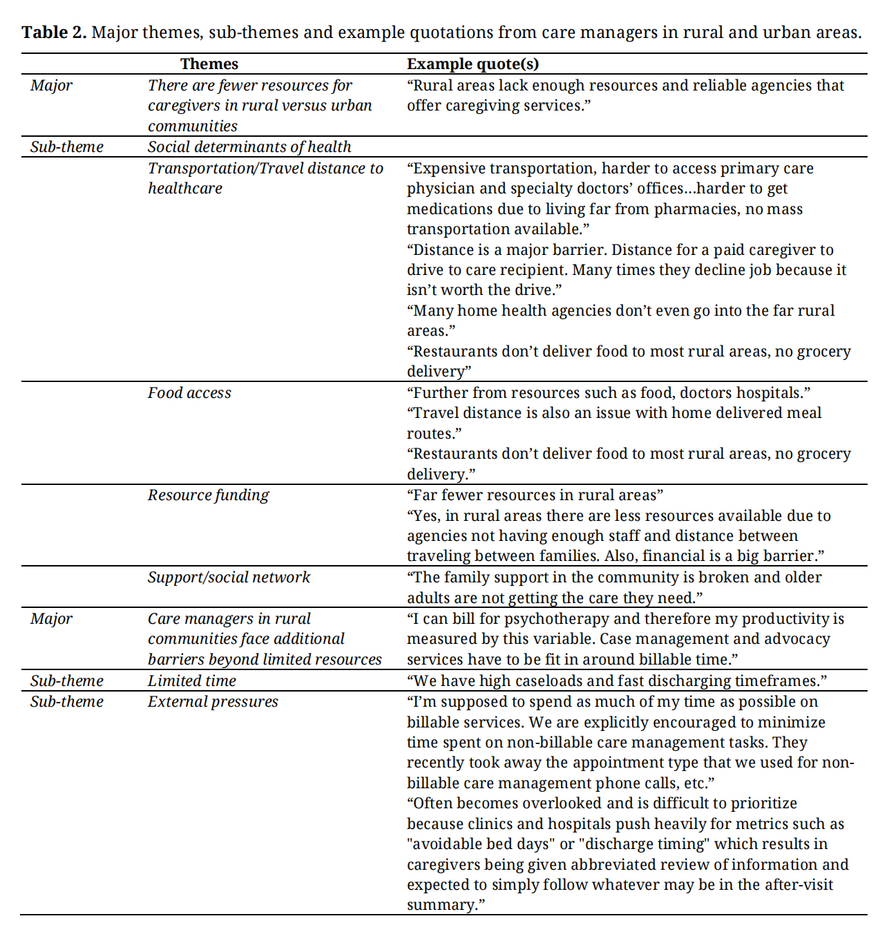 Table 2. Major themes, sub-themes and example quotations from care managers in rural and urban areas.
Overall, care managers practicing in rural and urban settings reported largely similar caregiver needs; however, financial need emerged as a notable exception, with rural practitioners more likely than urban practitioners to identify it as an important concern for caregivers. Our exploratory study provides insight into the perceived needs of informal caregivers from the perspective of care managers and suggests that geographic differences may be less pronounced than differences in resource availability and accessibility. While limited by sample size, our qualitative responses overwhelmingly showed that, regardless of geographical practice location, rural areas were perceived by care managers to have more barriers for caregiver support based on unique SDOH to rural areas and external job pressures of the care manager that limit the necessary time to find resources for rural caregivers. Having a better understanding of caregiver needs based on geographical location and location of existing resources will help improve support for caregivers of older adults in rural areas, as well as the practitioners who assist them.
Due to our small sample size, conclusions from group comparisons should be interpreted with caution. However, because of the exploratory nature of the study, it is important to expand upon the similarities and differences between groups. Across settings, care managers emphasized that resource limitations adversely affect caregiver support, particularly in the domains of financial need, food security, and healthcare access, similar to domains highlighted in Douthit et al. (2015) [16] and Matthews et al. (2025) [17]. Although rural and urban care managers reported largely similar perceptions of informal caregiver needs, these findings underscore the importance of validating care manager perspectives with caregivers themselves, as motivations for utilizing services may differ by geographic context. Prior work by Miyawaki et al. (2024) [18] showed that rural, informal caregivers may experience guilt related to using available resources, which may influence service uptake. Consistent with this, our findings showed care managers serving rural areas placed significantly greater emphasis on the financial needs of rural caregivers. This is consistent with Bouldin et al.’s observation that rural, informal caregivers experience higher financial strain than their urban counterparts [19]. The relationship between caregiving demands and perceived burden is complex and heterogeneous, as caregivers’ intrinsic beliefs may influence how caregiving experiences are interpreted and contribute to perceived burden and potentially, resource-utilization [20]. These observations suggest the need for further investigation with a larger sample to identify more specifically the factors contributing to higher financial needs of informal caregivers motivations for utilization.
In addition to the ecological factors impacting caregiver needs, we found that the time restraints and billing pressure on care managers are another potential barrier for providing adequate caregiver support [19]. Our participants, in both rural and urban areas, noted that already limited resources for caregivers in rural areas lead to the need for additional time to find resources. This ‘extra’ time does not exist within the current healthcare billing structure. These observations support the need to more specifically understand the needs of care managers to best support informal caregivers as well as optimize career satisfaction.
While most participants who correctly identified the rural status of their practice area as measured by ZIP-code rurality, nearly half did not. This observation is consistent with previous work that showed only moderate agreement between individuals’ self-identified rurality and standard Rural-Urban Commuting Areas (RUCA)-based, indicating that people may feel misclassified by conventional definitions of rurality [21]. The ambiguity of the definition of rurality suggests the complexity of fully understanding the ecological factors that may influence the relationship between geographical residence and caregiver burden. We observed this in our sensitivity analysis using self-reported rural status, in which care managers perceived that rural caregivers had more requests, and financial concerns were no longer significantly associated with rural residence as they were in the verified geographical group. Notably, we chose to use the “Am I Rural” definition of rurality, which uses county-level data [13]. As seen in work by Zahnd et al., the variable definitions used to define rural can impact interpretation of care access and perpetuate unmet needs unintentionally [22]. Therefore, standardizing the definition of urban and rural practice location could potentially eliminate any inconsistencies when conducting research based on rural and urban geographical differences. This is relevant particularly for program funding, as resource funding eligibility can be based on different criteria set by organizations such as, the United States Department of Agriculture criteria or Health Resources and Services Administration.
Strengths of this study include exploring practitioners’ perspectives of an under-studied group with unique unmet needs. However, this work presents limitations, such as the small sample size and use of convenience sampling from certain departments that were known to have social workers and care coordinators. The sensitivity analysis comparing self-reported versus actual geographic practice area was conducted to strengthen the analytical approach despite the small sample size. Because demographic information was inconsistently available for non-respondents due to use of mass email accounts (e.g., listservs), we were not able to evaluate differences between responders and non-responders. Therefore, the possibility of nonresponse bias should be considered when interpreting the findings. Additionally, focus of the study to North Carolina specifically limits the external validity of this study to caregivers in other parts of the country. Perceptions of rurality might also vary within North Carolina compared to other states because of the high proportion of rural residents. North Carolina is known to have a high rural population second only to Texas [23]. While NC is experiencing increasing urbanization, a large portion of its counties still retain rural character [24]. Lastly, there is no gold standard definition for rurality, for which our definition may over- or underestimate patterns of caregiving needs.
Care managers practicing in rural and urban areas report unmet needs of caregivers in rural areas related to poor resource access and competing pressures to best support rural, informal caregivers. Care managers thought unmet needs were driven by social determinants of health and believed that system-level pressures present additional barriers. These findings are important to guide future work examining the necessary support required for care managers as they provide support for informal caregivers of older adults in under-resourced areas.
This was considered an exempt study by the University of North Carolina-Chapel Hill Institutional Review Board (IRB #24-3130). All participants opted in to survey participation for this exempt study.
Declaration of Helsinki STROBE Reporting GuidelineThis study adhered to the Helsinki Declaration. The Strengthening the Reporting of Observational studies in Epidemiology (STROBE) reporting guideline were followed.
The following supplementary materials are available online, Table S1: Survey questions assessing the needs of informal caregivers in rural versus urban areas from the perspective of care managers; Table S2: Reported Types and Frequency of Care Management Support Provided to Informal Caregivers Across Rural and Urban Practices; Table S3: Reported Types and Frequency of Care Management Support Provided to Informal Caregivers Across Self-Reported Rural and Urban Practices.
Data is available upon request.
HS, ND participated in the conceptualization, analysis, writing, and critical review of the manuscript. BG, JM, TT, DD participated in conceptualization, data interpretation, writing, and critical review of the manuscript.
The authors declare that they have no conflicts of interest.
This project was not supported by any funding.
1.
2.
3.
4.
5.
6.
7.
8.
9.
10.
11.
12.
13.
14.
15.
16.
17.
18.
19.
20.
21.
22.
23.
24.
Spangler H, Daneshvar N, Godfrey B, Muise J, Thompson T, Dews D. Unmet Informal Caregiver Needs in Rural Areas: A Care Management Perspective. Adv Geriatr Med Res. 2026;8(3):e260019. https://doi.org/10.20900/agmr20260019.

Copyright © Hapres Co., Ltd. Privacy Policy | Terms and Conditions


